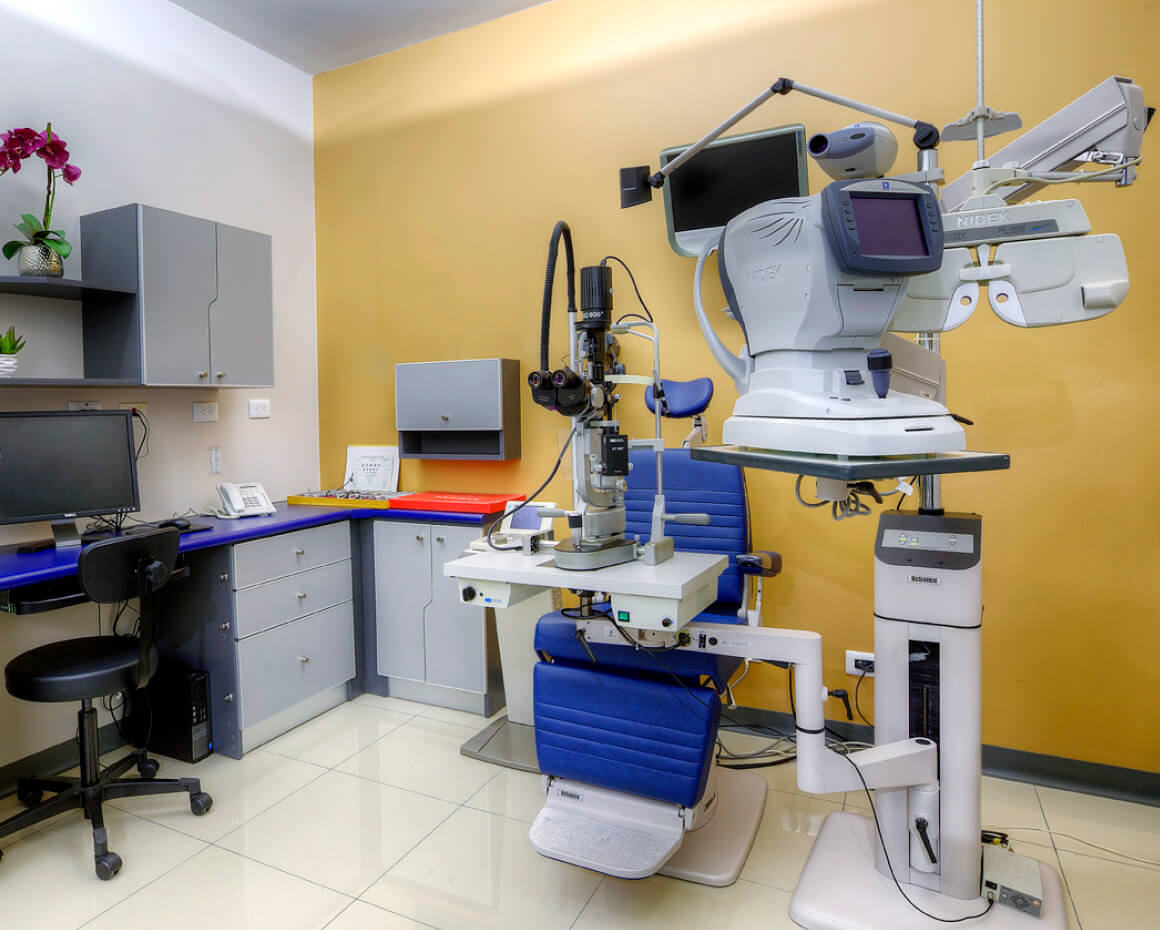
Our Clinic
Our specialized staff and state-of-the-art technology are completely at your service.
Our Services
We offer specialized and comprehensive services for the prevention, diagnosis, treatment and control of eye diseases. Our team of ophthalmologists, with different subspecialties and recognized experience, are at your service, creating health and wellness so that you continue to see the life you want and deserve to see.
Clinical Studies:
Special Exams
These are the diagnostic tests we perform that are essential to:
- make a correct diagnosis
- indicate the optimal treatment for each patient
All these tests:
- require a prior appointment. However, if the day of your medical consultation the doctor requires you to get a special test for his diagnosis, it will be performed right away.
We will gladly help you to have your test done as soon as possible.
Learn more by clicking on each one of them!
Angiography
Fluorescein angiography is a diagnostic test that is used to study the vascular circulation of the retina and, to a lesser [...]
View moreAngiography
Fluorescein angiography is a diagnostic test that is used to study the vascular circulation of the retina and, to a lesser extent, the layer that is under it and that nourishes it, the choroid. Although they are highly vascularized tissues, their networks of blood vessels cannot be seen with the naked eye.Hence, it is necessary to carry out this test, which despite the appearance of less invasive options is still very useful due to its high sensitivity when evaluating, quantifying and locating retinal lesions.
What does it consist of?
Fluorescein angiography consists of the intravenous injection of a contrast substance that travels through the blood to reach the blood vessels in the back of the eye and stain them. In this way, it allows them to be viewed by using sophisticated photographic equipment, which captures the images to form a complete map of the ocular vascular network.The patient requires pupil dilation, it is recommended to come with a companion. The patient should avoid drinking coffee, tea or soft drinks and coming on an empty stomach.
1
hour
time to perform this test
Indicated for patients with:
- Diabetic retinopathy
- Diabetic macular edema
- Macular degeneration
- Central serous retinopathy
- Macular holes
- Epiretinal membranes
- Degenerative diseases of the retina such as retinitis pigmentosa
- Eye tumors
Angio-OCT
Angio-OCT is a new diagnostic equipment that uses optical coherence tomography (OCT) technology, to obtain [...]
View moreAngio-OCT
Angio-OCT is a new diagnostic equipment that uses optical coherence tomography (OCT) technology, to obtain high-quality images of the retinal circulation, in addition to visualizing the structures of the posterior pole of the eye in 3D. In this way, it offers the opportunity to examine with extreme precision the vascular network of the eye at different depths, showing the mobility of the circulation in the blood vessels in the specific layers of the retina and the choroid (layer that is just below) , as well as in the head of the optic nerve.What does it consist of?
This technique, based on OCT (light emission) technology, captures images in just 3 seconds. It is quick to carry out and also avoids discomfort and possible adverse effects on patients. This makes it a minimally invasive option that can be repeated more frequently for comprehensive disease control. OCT angiography is performed at consultation appointments in less than 10 minutes, without requiring previous preparation for the patient or dilation of the pupil.Painless patient test.
It can be done without prior preparation, and without pupillary dilation.
Can be performed on patients of any age.
A device is used that emits light into the eye through the pupil, allowing the different layers of the retina and choroid to be analyzed.
It is a minimally invasive test for the patient.
-10
minutes
time to perform this test
La angio-OCT se realiza en consulta, sin requerir preparación previa del paciente ni dilatación de la pupila.
It is indicated in cases of:
- Macular hole
- MHigh myopia
- AMD
- Stargardt disease
- Glaucoma
- Macular Epiretinal Membrane (MEM)
- Venous retinal occlusions
- Diabetic retinopathy
- Retinitis pigmentosa
- Intraocular tumors
- Uveítis
Eye fundus photographs
Multicolor or black and white eye fundus photographs: series of photographs of the inner part of the eyeball that document [...]
View moreMulticolor or black and white eye fundus photographs
series of photographs of the inner part of the eyeball that document the different diseases and monitor their progression over time. The patients may have their pupils dilated.
10
minutes
estimated time for this test
Indicated for patients with:
- Conjunctival nevus
- Cataract
- Intraocular lens subluxation
- Macular alterations, scars or diseases of the optic nerve
Biometry
Measurement of several ocular dimensions used for calculation of intraocular lens power for cataract surgery, that uses [...]
View moreBiometry
Measurement of several ocular dimensions used for calculation of intraocular lens power for cataract surgery, that uses two systems: using light with laser interferometry (Lenstar®) or ultrasound (ultrasonic). This is a diagnostic special study that allows measuring the size of the eye from the outermost part to the deepest part, and that is used to calculate intraocular lenses for cataract surgery or Phaco refractive surgery.
It is very important to point out that for this examination the patient should have discontinued the use of contact lenses for several days. Ideally, the patient should not have his pupils dilated.
15
minutes
time to perform this test
Corneal Topography (Pentacam®)
It is a non-invasive imaging technique, to map the curvature, thickness and several other [...]
View moreCorneal Topography (Pentacam®)
It is a non-invasive imaging technique, to map the curvature, thickness and several other characteristics of the cornea. A topographer is an instrument that allows us to evaluate the cornea in detail through the visualization of different color maps in which the curvature, thickness, elevation and optical aberrations of the cornea are displayed. Pentacam is the brand of the instrument we use. It actually works as tomography because, besides corneal evaluation, we can also use it to evaluate a great part of the anterior segment of the eye.
Preparation of the patient
It should not be done if the patient has recently worn contact lenses, because this might change the corneal surface causing measurement errors. If the patient uses soft contact lenses, the patient must stop their use for a minimum of 3 to 5 days, and with rigid lenses the patient must suspend their use at least 8 days before the appointment. Ideally, the patient is not dilated.15
minutes
time to perform this test
It is referred in cases of:
- Diagnosis of different corneal pathologies
- Approach to cataract or corneal surgery
- LASIK refractive surgery
- Keratoconus diagnosis and follow up
- Contact lenses fitting
Pachymetry
It is an ophthalmological test used to measure the thickness of the cornea, the transparent lens located at the front of the eye.
View morePachymetry
It is an ophthalmological test used to measure the thickness of the cornea, the transparent lens located at the front of the eye.
10
minutes
time to perform this test
It is referred in cases of:
- Diseases of the cornea
- Prior to refractive surgery
- Presumed glaucoma
Specular microscopy
Test that evaluates the quantity and quality of corneal endothelial cells to assess the health status of the cornea.
View moreSpecular microscopy
Test that evaluates the quantity and quality of corneal endothelial cells to assess the health status of the cornea.
10
minutes
time to perform this test
It is referred in cases of:
- Corneal dystrophies
- Corneal trauma
- Corneal transplant rejection
- Unexplained corneal edema
- Recurrent epithelial erosions
- Cataract surgery preoperative evaluation
- Phakic intraocular lens (Visian ICL) implantation and follow up
O.R.A
Study of corneal hysteresis, which is the factor of flexibility and corneal resistance. It is a corneal biomechanical [...]
View moreO.R.A
Study of corneal hysteresis, which is the factor of flexibility and corneal resistance. It is a corneal biomechanical evaluation performed with an air pulse applied to the cornea, a light-emitting diode, and a receiver. Anesthetic eye drops are placed to avoid discomfort. It is a painless test.
15
minutes
time to perform this test
It is referred in cases of:
- Postoperative ectasia
- Intraocular pressure evaluation
- Keratoconus diagnosis and follow up
Computerized campimetry
Campimetry or Visual Field Perimetry is an ophthalmological test that measures the total area in which we are able [...]
View moreComputerized campimetry
Campimetry or Visual Field Perimetry is an ophthalmological test that measures the total area in which we are able to see objects around a fixed point. In this sense, it allows us to determine the amplitude of our peripheral or lateral vision, once we have fixed our gaze on an object.
Likewise, it is used to study and measure the evolution of the visual field from time to time or periodically.
What does it consist of?
It is a test that is done in a room with very little light or in the dark. It is an easy test but requires high collaboration from the patient.The perimeter emits small moving light points of different intensities (static or computerized perimetry).
The patient sits and rests his head on this machine that sends small light points or flashes. The goal is for the patient to recognize peripheral lights, without looking away from the center point. When viewing each light, the patient must press a button.
As the test progresses, the perimeter draws a vision map, showing the areas that the patient is able to see and those that are not.
1
hour
time to perform this test
It is referred in cases of:
- Glaucoma follow up
- Appearance of the abnormal optic nerve
- Evaluation of eyelid surgery
- Suspicion of brain tumors and neurological disorders
OCT
It is a non-invasive optical tomographic imaging (section imaging) technique that offers high-resolution photographs [...]
View moreOCT
It is a non-invasive optical tomographic imaging (section imaging) technique that offers high-resolution photographs of the different layers of the retina and the optic nerve. The acronym stands for "Optical Coherence Tomography." It is an exam that allows us to assess the innermost cell layers of the eye, especially those of the macula, which is the central and most important part of the retina, and it also allows us to assess and quantify the fibers of the optic nerve. Anterior Segment OCT can also be performed, in which we can evaluate the anterior structures of the eye, such as the cornea, anterior chamber and crystalline lens.
15
minutes
time to perform this test
It is referred in cases of:
- Retinitis pigmentosa
- Diabetic macular edema
- Venous occlusions
- Central serous choroidopathy
- Macular hole
- Macular epiretinal membrane
- Diabetic retinopathy
- Dry or atrophic AMD
- Exudative or wet AMD
- Pathological myopia
- Retinal detachment
- Vitreomacular traction syndrome
- Subretinal hemorrhages
- Proliferative diabetic retinopathy
- Glaucoma
- Phakic intraocular lens implantation
Blue peak
Non-invasive scanning modality that reveals the metabolic stress of the retina using lipofuscin as an indicator. [...]
View moreBlue peak
Non-invasive scanning modality that reveals the metabolic stress of the retina using lipofuscin as an indicator. The images can reveal diseases in the retinal pigment epithelium and malfunctions in receptor cells offering diagnostic perspectives in retinal conditions such as AMD and inherited diseases. The patient needs to be dilated.
10
minutes
time to perform this test
Angiography
Fluorescein angiography is a diagnostic test that is used to study the vascular circulation of the retina and, to a lesser extent [...]
View MoreAngiography
Fluorescein angiography is a diagnostic test that is used to study the vascular circulation of the retina and, to a lesser extent, the layer that is under it and that nourishes it, the choroid. Although they are highly vascularized tissues, their networks of blood vessels cannot be seen with the naked eye.Hence, it is necessary to carry out this test, which despite the appearance of less invasive options is still very useful due to its high sensitivity when evaluating, quantifying and locating retinal lesions.
What does it consist of?
Fluorescein angiography consists of the intravenous injection of a contrast substance that travels through the blood to reach the blood vessels in the back of the eye and stain them. In this way, it allows them to be viewed by using sophisticated photographic equipment, which captures the images to form a complete map of the ocular vascular network.The patient requires pupil dilation, it is recommended to come with a companion. The patient should avoid drinking coffee, tea or soft drinks and coming on an empty stomach.
1
hour
approximately time to perform this test
Indicated for patients with:
- Diabetic retinopathy
- Diabetic macular edema
- Macular degeneration
- Central serous retinopathy
- Macular holes
- Epiretinal membranes
- Degenerative diseases of the retina such as retinitis pigmentosa
- Eye tumors
Angio-OCT
Angio-OCT is a new diagnostic equipment that uses optical coherence tomography (OCT) technology, to obtain [...]
View moreAngio-OCT
Angio-OCT is a new diagnostic equipment that uses optical coherence tomography (OCT) technology, to obtain high-quality images of the retinal circulation, in addition to visualizing the structures of the posterior pole of the eye in 3D. In this way, it offers the opportunity to examine with extreme precision the vascular network of the eye at different depths, showing the mobility of the circulation in the blood vessels in the specific layers of the retina and the choroid (layer that is just below) , as well as in the head of the optic nerve.What does it consist of?
This technique, based on OCT (light emission) technology, captures images in just 3 seconds. It is quick to carry out and also avoids discomfort and possible adverse effects on patients. This makes it a minimally invasive option that can be repeated more frequently for comprehensive disease control. OCT angiography is performed at consultation appointments in less than 10 minutes, without requiring previous preparation for the patient or dilation of the pupil.Painless patient test.
It can be done without prior preparation, and without pupillary dilation.
Can be performed on patients of any age.
A device is used that emits light into the eye through the pupil, allowing the different layers of the retina and choroid to be analyzed.
It is a minimally invasive test for the patient.
-10
minutes
time to perform this test
La angio-OCT se realiza en consulta, sin requerir preparación previa del paciente ni dilatación de la pupila.
It is indicated in cases of:
- Macular hole
- MHigh myopia
- AMD
- Stargardt disease
- Glaucoma
- Macular Epiretinal Membrane (MEM)
- Venous retinal occlusions
- Diabetic retinopathy
- Retinitis pigmentosa
- Intraocular tumors
- Uveítis
Eye fundus photographs
Multicolor or black and white eye fundus photographs: series of photographs of the inner part of the eyeball that document [...]
View moreMulticolor or black and white eye fundus photographs
series of photographs of the inner part of the eyeball that document the different diseases and monitor their progression over time. The patients may have their pupils dilated.
10
minutes
estimated time for this test
Indicated for patients with:
- Conjunctival nevus
- Cataract
- Intraocular lens subluxation
- Macular alterations, scars or diseases of the optic nerve
Biometry
Measurement of several ocular dimensions used for calculation of intraocular lens power for cataract surgery, that uses [...]
View moreBiometry
Measurement of several ocular dimensions used for calculation of intraocular lens power for cataract surgery, that uses two systems: using light with laser interferometry (Lenstar®) or ultrasound (ultrasonic). This is a diagnostic special study that allows measuring the size of the eye from the outermost part to the deepest part, and that is used to calculate intraocular lenses for cataract surgery or Phaco refractive surgery.
It is very important to point out that for this examination the patient should have discontinued the use of contact lenses for several days. Ideally, the patient should not have his pupils dilated.
15
minutes
time to perform this test
Corneal Topography (Pentacam®)
It is a non-invasive imaging technique, to map the curvature, thickness and several other [...]
View moreCorneal Topography (Pentacam®)
It is a non-invasive imaging technique, to map the curvature, thickness and several other characteristics of the cornea. A topographer is an instrument that allows us to evaluate the cornea in detail through the visualization of different color maps in which the curvature, thickness, elevation and optical aberrations of the cornea are displayed. Pentacam is the brand of the instrument we use. It actually works as tomography because, besides corneal evaluation, we can also use it to evaluate a great part of the anterior segment of the eye.
Preparation of the patient
It should not be done if the patient has recently worn contact lenses, because this might change the corneal surface causing measurement errors. If the patient uses soft contact lenses, the patient must stop their use for a minimum of 3 to 5 days, and with rigid lenses the patient must suspend their use at least 8 days before the appointment. Ideally, the patient is not dilated.15
minutes
time to perform this test
It is referred in cases of:
- Diagnosis of different corneal pathologies
- Approach to cataract or corneal surgery
- LASIK refractive surgery
- Keratoconus diagnosis and follow up
- Contact lenses fitting
Pachymetry
It is an ophthalmological test used to measure the thickness of the cornea, the transparent lens located at the front of the eye.
View morePachymetry
It is an ophthalmological test used to measure the thickness of the cornea, the transparent lens located at the front of the eye.
10
minutes
time to perform this test
It is referred in cases of:
- Diseases of the cornea
- Prior to refractive surgery
- Presumed glaucoma
Specular microscopy
Test that evaluates the quantity and quality of corneal endothelial cells to assess the health status of the cornea.
View moreSpecular microscopy
Test that evaluates the quantity and quality of corneal endothelial cells to assess the health status of the cornea.
10
minutes
time to perform this test
It is referred in cases of:
- Corneal dystrophies
- Corneal trauma
- Corneal transplant rejection
- Unexplained corneal edema
- Recurrent epithelial erosions
- Cataract surgery preoperative evaluation
- Phakic intraocular lens (Visian ICL) implantation and follow up
O.R.A
Study of corneal hysteresis, which is the factor of flexibility and corneal resistance. It is a corneal biomechanical [...]
View moreO.R.A
Study of corneal hysteresis, which is the factor of flexibility and corneal resistance. It is a corneal biomechanical evaluation performed with an air pulse applied to the cornea, a light-emitting diode, and a receiver. Anesthetic eye drops are placed to avoid discomfort. It is a painless test.
15
minutes
time to perform this test
It is referred in cases of:
- Postoperative ectasia
- Intraocular pressure evaluation
- Keratoconus diagnosis and follow up
Computerized campimetry
Campimetry or Visual Field Perimetry is an ophthalmological test that measures the total area in which we are able [...]
View moreComputerized campimetry
Campimetry or Visual Field Perimetry is an ophthalmological test that measures the total area in which we are able to see objects around a fixed point. In this sense, it allows us to determine the amplitude of our peripheral or lateral vision, once we have fixed our gaze on an object.
Likewise, it is used to study and measure the evolution of the visual field from time to time or periodically.
What does it consist of?
It is a test that is done in a room with very little light or in the dark. It is an easy test but requires high collaboration from the patient.The perimeter emits small moving light points of different intensities (static or computerized perimetry).
The patient sits and rests his head on this machine that sends small light points or flashes. The goal is for the patient to recognize peripheral lights, without looking away from the center point. When viewing each light, the patient must press a button.
As the test progresses, the perimeter draws a vision map, showing the areas that the patient is able to see and those that are not.
1
hour
time to perform this test
It is referred in cases of:
- Glaucoma follow up
- Appearance of the abnormal optic nerve
- Evaluation of eyelid surgery
- Suspicion of brain tumors and neurological disorders
OCT
It is a non-invasive optical tomographic imaging (section imaging) technique that offers high-resolution photographs [...]
View moreOCT
It is a non-invasive optical tomographic imaging (section imaging) technique that offers high-resolution photographs of the different layers of the retina and the optic nerve. The acronym stands for "Optical Coherence Tomography." It is an exam that allows us to assess the innermost cell layers of the eye, especially those of the macula, which is the central and most important part of the retina, and it also allows us to assess and quantify the fibers of the optic nerve. Anterior Segment OCT can also be performed, in which we can evaluate the anterior structures of the eye, such as the cornea, anterior chamber and crystalline lens.
15
minutes
time to perform this test
It is referred in cases of:
- Retinitis pigmentosa
- Diabetic macular edema
- Venous occlusions
- Central serous choroidopathy
- Macular hole
- Macular epiretinal membrane
- Diabetic retinopathy
- Dry or atrophic AMD
- Exudative or wet AMD
- Pathological myopia
- Retinal detachment
- Vitreomacular traction syndrome
- Subretinal hemorrhages
- Proliferative diabetic retinopathy
- Glaucoma
- Phakic intraocular lens implantation
Blue peak
Non-invasive scanning modality that reveals the metabolic stress of the retina using lipofuscin as an indicator. [...]
View moreBlue peak
Non-invasive scanning modality that reveals the metabolic stress of the retina using lipofuscin as an indicator. The images can reveal diseases in the retinal pigment epithelium and malfunctions in receptor cells offering diagnostic perspectives in retinal conditions such as AMD and inherited diseases. The patient needs to be dilated.
10
minutes
time to perform this test
Our treatments and surgical procedures
We offer you the most innovative treatments and surgical techniques which exist worldwide, to correct and treat your visual problems.
Learn more by clicking on each one of them!
The 8 answers you should know
- 1
- What is a cataract?
A cataract is any opacity that develops in the crystalline lens, the natural lens inside the eye, behind the iris. The most frequent cataract is the one that occurs in relation to the aging process, but there are many other causes like ocular trauma, the use of some types of medications, inflammatory processes of the eye, genetic causes, among others. - 2
- To which patients is this procedure recommended?
To patients who show a slight, moderate or severe visual loss produced by an opacity in the natural lens of the eye. This type of surgery can also be indicated for patients over 50 years of age who, although they do not have a cataract yet, wish to correct their myopia, astigmatism, hyperopia or presbyopia to become independent from their glasses or contact lenses: this is what is known as Phacorrefractive surgery or Clear Lens Exchange. - 3
- What are the basic steps of this surgery?
The entire procedure is performed through a small 2mm incision on the periphery of the cornea. A technique called Phacoemulsification is used, which works with a system based on ultrasound, irrigation and aspiration to dissolve and aspirate the entire lens, leaving only the capsule or bag that surrounds it, which is then left completely empty. Then, immediately, an intraocular lens is implanted and will be placed inside that same bag that surrounded the natural lens. - 4
- What types of intraocular lenses are there?
The technological evolution in intraocular lenses in the last decades has been impressive. Before the surgery, special studies are carried out to determine the power of the intraocular lens that is needed for each eye. With current equipment, these measurements are very accurate, with very low error ranges. We currently use the following types of intraocular lenses:
- Monofocal IOL: with this type of lens, all the light that enters the eye is brought to a single focal point. In most cases the power of the lens is calculated so that the person has excellent distance vision without the need for glasses and only needing glasses for near reading.
- Toric monofocal IOL: this has the same characteristics mentioned for the monofocal lens, but it is a lens that additionally corrects astigmatism. Astigmatism is when the cornea has a meridian with a greater curvature and power than the other meridian, generating several different focal lines in the eye instead of a single focal point. This type of intraocular lenses then have different powers in their axes to compensate for this situation, thus correcting astigmatism, and giving a better visual result.
- Multifocal IOL: in this group there are several different technologies that allow the intraocular lens to divide light that enters the eye towards different focal points, so that the person obtains good visual acuity at all distances. In this way, the patient becomes independent from glasses for the vast majority of her life activities. Each case must be evaluated individually to determine if the person is a good candidate for a multifocal lens.
- Multifocal toric lens: This lens brings together the features mentioned above for multifocal lenses and toric lenses in a single lens.
- 5
- What can a patient expect from this surgery?
The surgery takes around 20 minutes per eye, depending on the case, and it is completely an outpatient procedure, that is, the patient goes home as soon as the surgery is done. In the vast majority of cases, the procedure is not performed on both eyes on the same day, but each eye is done on separate days. In most cases, only local anesthesia is used in eye, but if the patient wishes, mild sedation can be given. In the vast majority of cases , vision is already quite good the day after the procedure , although it will continue to improve in the next following days. - 6
- What are the benefits of cataract surgery?
It is a safe and fairly accurate method with extremely low risk for complications. The patient's vision is completely restored. It is a procedure that gives a definitive solution to the cataract. That is, it is impossible for it to come back. Cataract surgery can also correct both myopia and astigmatism, hyperopia and presbyopia. Extremely high refractive errors that cannot be treated with laser techniques can be corrected with intraocular lenses. The recovery process is fast. Visual acuity is already quite good the day after surgery and improves even more during the first week. - 7
- These are the steps that the patient interested in cataract surgery must follow:
Step 1. You should schedule an initial assessment consultation.
These are the tests that you will have on your first appointment:
Complete ophthalmological exam:- Optometric exam.
- Slit lamp biomicroscopy.
- Eye fundus exam (with dilated pupils).
- Eye pressure measurement.
- Individual diagnostic consultation with the ophthalmologist.
Step 2. If the doctor in the evaluation consultation confirms that the patient is indeed a candidate for cataract or phacorefractive surgery, he will proceed to indicate to the patient that he should carry out some studies to determine if he is a good candidate as well as calculating the power and type of lens to be implanted in each eye: 1. Corneal Topography / Tomography, 2. Specular Microscopy (corneal endothelial cells evaluation) and 3. Optical Biometry (measurement of internal dimensions of the eye) to calculate the power of the IOL. - 8
- It is very important to stop wearing contact lenses 1 week before the evaluation appointment for the studies to be reliable.
The 6 answers you should know
- 1
- Which patients are recommended to have this surgery?
To patients who want to correct their myopia, astigmatism and / or hyperopia to become independent from their glasses or contact lenses. In some cases to patients with presbyopia. Generally, patients over 20 years of age can be operated on, because that is when the eye has finished growing and the refractive errors are stable. - 2
- What is LASIK?
Laser Assisted in Situ Keratomileusis is a procedure performed with an Excimer laser equipment with which a photoablation is performed on the cornea, thus changing its curvature and optical power. With this modification of the cornea, any refractive defect that the person has is corrected. In the procedure, a very precise cut is first made, creating a thin, superficial layer on the cornea (corneal flap), which is lifted for a few seconds. At this moment the laser platform performs the treatment on the underlying corneal tissue changing its curvature and then, immediately, this thin layer is repositioned adhering by itself again. - 3
- What is the difference between LASIK and FemtoLASIK?
In the first part of LASIK surgery, a very precise cut is made creating a corneal flap which is lifted so that the curvature and power correction of the cornea can then be carried out with the Excimer laser. Then this cap or flap is repositioned and adheres on its own again. The creation of this layer or corneal flap can be done with an automated mechanical knife (microkeratome) as it is done in classic LASIK, or more recently, with a laser that allows very precise cuts called the Femtosecond Laser. So in the FemtoLASIK, the difference is that this initial cut is made with the laser, making it even safer and more precise. - 4
- What can a patient expect from this surgery?
The surgery is very fast, it can last around 7 to 10 minutes per eye, depending on the case. It is performed under topical anesthesia (drops) on the eye only. The precision and safety of this surgery makes it the surgical procedure of choice for most refractive errors. Recovery is very fast, even showing a quite good visual acuity the very first day after surgery, although this will improve during the next days following the procedure. Currently it is one of the safest and most effective techniques for the correction of visual defects. - 5
- What are the benefits of LASIK surgery?
- It is a safe and accurate method.
- Very low risk of infection.
- Myopia, astigmatism and hyperopia can all be corrected.
- It is a painless procedure.
- Visual recovery is fast.
- The recovery process is quite easy. The use of electronic devices, computer, television, reading, etc., can be done even the day after the procedure. However, you do have to avoid sports that involve risk of blows to the face or eye exposure to pools and sea water.
- 6
- These are the steps that the patient interested in LASIK surgery should follow:
Step 1. You should schedule an initial assessment consultation.
These are the tests that you will have on your first appointment:
Complete ophthalmological exam:
- Optometric exam.
- Slit lamp biomicroscopy.
- Fundus exam (with dilated pupils).
- TEye pressure measurement.
- Individual diagnostic consultation with the ophthalmologist.
- 7
- It is very important to stop the use of contact lenses 1 week before the evaluation appointment so that the Corneal Topography study is reliable.
The 6 answers you should know
- 1
- Which patients are recommended to have this surgery?
Patients who want to correct their myopia, astigmatism and / or hyperopia to become independent from their glasses or contact lenses. In some cases patients with presbyopia. Generally, patients over 20 to 23 years of age are the ones that are candidates for these procedures because that is when the eye stops growing and the refractive errors are stable. - 2
- What is PRK?
Photorefractive Keractectomy is a procedure performed with an Excimer laser equipment with which a photoablation is performed on the cornea, thus changing its curvature and optical power. With this modification of the cornea, any refractive defect that the person has is corrected. In the procedure, an area of the outermost layer of the cornea, called the epithelium, is first removed. It is important to emphasize that no cut is made in the cornea, only that thin layer is removed, which will regenerate during the following days. After the removal of said epithelium, the laser performs the treatment on the corneal tissue by changing its curvature and then a protective contact lens is placed that will remain for about 3 days while that epithelial layer grows back again. - 3
- What can a patient expect from this surgery?
The surgery is very fast, it can last around 7 minutes per eye, depending on the case, and it is performed under topical anesthesia with drops on the eye. The final visual result is excellent, completely equivalent to that of other techniques such as LASIK and SMILE, but the recovery process is a bit slower and annoying since you have to wait a few days for the regeneration and stabilization of that layer called epithelium so that the vision becomes stable. There can be some pain, photofobia and discomfort during the first 3 days after surgery. However, this technique has the great advantage that there will be no cut or flap in the cornea and, as it was stated before, the visual result is exactly the same as in LASIK and SMILE. - 4
- What are the benefits of PRK surgery?
- It is a safe and accurate method.
- Myopia, astigmatism and hyperopia can all be corrected.
- No cut is made to the cornea, which in turn means no corneal flap, avoiding potential problems if there is some kind of trauma to the eye.
- Many patients with thinner corneas who are not candidates for LASIK can be operated on.
- The visual result is excellent, although it takes more days to fully recover.
- 5
- These are the steps that the patient interested in PRK surgery should follow:
Step 1. You should schedule an initial assessment consultation.
These are the tests that you will have on your first appointment:
Complete ophthalmological exam:
- Optometric exam.
- Slit lamp biomicroscopy.
- Fundus exam (with dilated pupils).
- Eye pressure measurement.
- Individual diagnostic consultation with the ophthalmologist.
- 6
- It is very important to stop the use of contact lenses 1 week before the evaluation appointment so that the Corneal Topography study is reliable.
The 6 answers you should know
- 1
- To which patients is recommended to have this surgery?
Patients who wish to correct their myopia and / or astigmatism. Generally, patients over 20 years of age are operated on, because that is when the eye has stopped growing and refractive errors are stable. - 2
- What is SMILE?
SMILE stands for Small Incision Lenticule Extraction. It is another one of the most advanced methods of corneal laser surgery for the correction of myopia and astigmatism. A laser platform called Femtosecond Laser is used. This laser is able to perform cuts within the cornea with an incredible precision of very few microns. Thanks to this precision, in SMILE, a cut is made within the corneal thickness creating a lenticule that will then be extracted through a small 2.5 mm peripheral incision. In this way, the curvature and power of the cornea are modified, correcting the person's refractive defect. - 3
- What can a patient expect from this surgery?
It is a safe, effective and less invasive surgery, with an intervention lasting approximately 5 to 10 minutes per eye. Only topical anesthesia in drops is used. The main difference and advantage of this technique, compared to its predecessors, is that the incision made in the cornea is only 2.5 mm and no corneal layer is lifted during the procedure. That is to say: there is no corneal flap. The advantage of this is that the risk of the corneal flap movement or dislocation due to accidental trauma in the first postoperative days is eliminated and on the other hand, the development of dry eye after surgery may be minimized. - 4
- What are the benefits of SMILE surgery?
The corneal lenticule is removed through a very small incision. Since the alteration on the corneal surface is so minimal, the development of long-term dry eye is less frequent and decreases with SMILE compared to LASIK. Some additional benefits are:- It is a safe and accurate method.
- The surgery is painless.
- Visual recovery is fast. The next day visual acuity is already quite good, but it improves during the first postoperative weeks.
- The recovery period is relatively quick. The use of electronic devices, computer, television, reading, etc., can be done as early as the day after the procedure. You can return to all sports activity almost immediately, except for swimming, for which it is preferred to wait 2 weeks after surgery.
- 5
- These are the steps that the patient interested in SMILE surgery must follow:
Step 1. You should schedule an initial assessment consultation.
These are the tests that you will have on your first appointment:
Complete ophthalmological exam:
- Optometric exam.
- Slit lamp biomicroscopy.
- Fundus exam (with dilated pupils).
- Eye pressure measurement.
- Individual diagnostic consultation with the ophthalmologist.
- 6
- It is very important to stop the use of contact lenses 1 week before the evaluation appointment so that the Corneal Topography study is reliable.
All the details are exactly the same as the ones for cataract surgery. You can read about all the important information regarding this procedure in the Cataract Surgery section.
The 6 answers you should know
- 1
- To which patients is recommended?
To patients who want to correct their myopia, astigmatism or hyperopia to become independent from their glasses or contact lenses. Generally, patients over 20 years of age are operated on, because that is when the eye has stopped growing and the refractive errors are stable. Many patients who are not candidates for corneal laser surgery can be operated on with an ICL lens implant and thus solve their refractive problem. It is especially useful in cases of high and very high refractive errors, although it is indicated in any degree of myopia, astigmatism or hyperopia. - 2
- What is ICL?
Implantable Collamer Lens, currently known as EVO Visian ICL, is a phakic intraocular lens. That is, it is a lens that can be implanted inside the eye without touching or modifying the internal natural lens called the crystalline lens. To put it simply, it is like a very small soft contact lens that is implanted inside the eye to correct refractive errors from low to very high. The lens is ordered with the specific correction power for each eye, as well as the precise size for that eye. The entire procedure is performed through a very small incision of just 2.5 mm in the periphery of the cornea. - 3
- What can a patient expect from this surgery?
The surgery is quite fast, it can last around 15 to 20 minutes per eye, depending on the case. In the vast majority of cases, local anesthesia is used, with drops in the eye only. But, if the patient wishes, mild sedation can be given. The day after the procedure, vision is already very good, although it will continue to sharpen in the following days. - 4
- What are the benefits of EVO Visian ICL implant surgery?
- It is a safe and accurate method.
- Myopia, astigmatism and hyperopia can all be corrected.
- Extremely high refractive errors that cannot be treated with laser techniques can be corrected with this procedure.
- There is no cut or any treatment performed on the cornea or the crystalline lens, keeping the natural lenses of the eye intact and healthy without any modification.
- Many patients with thin corneas who are not candidates for LASIK, PRK or SMILE can be operated on.
- The procedure is potentially "reversible." That is, the lens can be explanted at any time and the eye returns to its original state.
- 5
- These are the steps that the patient interested in an EVO Visian ICL implant surgery should follow:
Step 1. You should schedule an initial assessment consultation.
These are the tests that you will have on your first appointment:
Complete ophthalmological exam:
- Optometric exam.
- Slit lamp biomicroscopy.
- Fundus exam (with dilated pupils).
- Eye pressure measurement.
- Individual diagnostic consultation with the ophthalmologist.
- 6
- It is very important to stop the use of contact lenses 1 week before the evaluation appointment for the studies to be reliable. Once the Visian ICL lenses are ordered, it takes approximately 2 weeks for the lenses to arrive at our clinic in Costa Rica.
The 6 answers you should know
- 1
- What is a Pterygium?
The conjunctiva is a semitransparent membrane that covers the sclera (white part of the eye) and the eyelids internally. The cornea is the outer clear lens in front of the eye. Pterygium is an abnormal growth of the conjunctiva in which it exceeds the normal limit with the cornea and then advances over it, forming that "membrane" or "skin" that partially covers it. It is often misnamed "external cataract," but this is a misnomer and one that tends to confuse people. - 2
- To which patients is recommended?
To patients who have a pterygium that is causing symptoms such as red eye, foreign body sensation and frequent eye irritation. Also the aesthetic appearance of this membrane on the eye may be an indication for surgery. When the pterygium is large it can negatively influence vision and this is another reason to indicate a surgical resolution. - 3
- What does the surgery consist of?
In pterygium surgery, the abnormal membrane will be resected, leaving the cornea intact and clean again. But additionally, what we call a conjunctiva graft is performed, which consists of taking healthy conjunctiva from the areas that are under the eyelids, in which the membrane is very healthy, and we are going to take a piece of it to place it in the area where the pterygium was removed. In this way we provide this area with healthy tissue again, greatly reducing the possibility that this pterygium will grow back again. - 4
- What can a patient expect from this surgery?
The surgery is usually done under local anesthesia and there is no pain at all. However, if the patient wishes, light sedation can be given. The procedure lasts approximately 25 to 30 minutes and at the end you can go home immediately. The day after the procedure and for a few days there may be a slight foreign body sensation and the eye may be a little red. However, the vision is normal and the patient can carry out his normal daily activities except for swimming and sports in which the eye can get hit. It is advisable to protect yourself from the sun for a few days.
- 5
- What are the benefits of Pterygium surgery?
- It is a very safe method.
- The cause of eye irritation is eliminated.
- Normality of the surface of the eye is restored.
- The recovery process is simple and does not limit most daily activities.
- The chance that the problem recurs is extremely low if the surgery is done properly.
- 6
- These are the steps that the patient interested in a Pterygium surgery should follow:
You should schedule an initial assessment consultation. These are the exams that you will have at your appointment, as part of your comprehensive eye exam:- Optometric exam.
- Slit lamp biomicroscopy.
- Fundus exam (with dilated pupils).
- Eye pressure measurement.
- Individual diagnostic consultation with the ophthalmologist.
The 6 answers you should know
- 1
- To which patients is it recommended?
To patients who present a moderate or severe visual acuity loss caused by an alteration in the transparency, curvature or structure of one or more layers of the cornea and that it is not possible to correct it by other less invasive methods. Among the diseases that may require a corneal transplant are: keratoconus, corneal scars and ulcers, decompensation of the cornea after cataract surgeries (Bullous Keratopathy), genetic diseases of the cornea (Corneal Dystrophies), among others. - 2
- What is the cornea?
The cornea is the clear lens in front of the eye. It is responsible for most of the optical power of the eye. It is composed of a special type of collagen and other proteins that, due to their characteristics and arrangement, allow the tissue to be transparent and allow the passage and focus of light towards the inner part of the eye.
- 3
- What types of corneal transplants are there?
Depending on the type of problem or alteration that the cornea presents in each patient, the type of transplant indicated may be different for each case. There are cases in which all the layers of the cornea are affected and in this case a complete replacement of the entire thickness must be made, while in other cases only one or some specific layers are affected and then only these layers must be replaced maintaining the rest of the original cornea.- Penetrating Transplantation (Penetrating Keratoplasty): In this procedure, the full thickness of the cornea will be completely eliminated in a central diameter determined by the surgeon in each case, and will be replaced by a full-thickness donor cornea. That is, all the layers of the cornea are being replaced. The new tissue must be sutured so that it adheres to the recipient eye and these sutures will be removed little by little during the months after the surgery.
- Anterior Lamellar Transplant: In this case, only the anterior layers of the cornea will be replaced, keeping the original posterior layers of each patient. The new tissue must be sutured so that it adheres to the recipient eye and these sutures will be removed little by little during the months after the surgery. The modality of this group of transplants that we do most frequently is called Deep Anterior Lamellar Keratoplasty (DALK).
- Posterior lamellar transplants (Endothelial Transplant): This type of transplant is indicated when the corneal damage is in the deeper layers, specifically in the so-called endothelium. The endothelial cells are responsible for keeping the hydration of the cornea at a low level, and then when these cells are lost and reach very low levels the cornea fills up with fluid and loses its transparency. In this type of transplant, we are going to replace only that inner endothelial layer, keeping all the rest of the original thickness of the patient's cornea. In these procedures, a small incision of only approximately 3 mm is made and there is no progressive removal of sutures as in other types of transplantation. Among the modalities in this group of transplants are Descemet Stripping Automated Endothelial Keratoplasty (DSAEK) and Descemet Membrane Endothelial Keratoplasty (DMEK).
- 4
- Where are donor corneas obtained from?
Corneas are obtained from people who die from whom their corneas are extracted and these are processed, studied and preserved in what is known as an Eye Bank. We are affiliated with Eye Banks in the United States of America (USA) and from there we import corneas for our surgeries. The entire process, as well as the medical and auxiliary personnel involved, are endorsed by the Transplant Secretariat of the Ministry of Health of Costa Rica. - 5
- What benefits does Corneal Transplant surgery provide?
- The patient's vision is restored.
- In most cases the risk of complications is low.
- For pathology of the endothelium of the cornea, endothelial transplants have a great advantage over penetrating transplants, since recovery time and the risk of corneal rejection are greatly reduced.
- 6
- These are the steps that the patient interested in a Corneal Transplant surgery should follow:
You should schedule an initial assessment consultation. These are the exams that you will have at your first appointment, as part of your comprehensive eye exam:
- Optometric exam.
- Slit lamp biomicroscopy.
- Fundus exam (with dilated pupils).
- Eye pressure measurement.
- Individual diagnostic consultation with the ophthalmologist.
- Optical clarity: a. Hemorrhages commonly seen in people with Diabetes and other vascular diseases. b.Inflammation of different ethiologies c.Viiral, bacterial or parasitic infections and d.Ocular trauma.
- In its structure, changing the semi-liquid quality of the vitreous to form membranes within it. These changes can cause membranes on the retina, more precisely on the macula (central area of the retina where central and most sharp vision is processed). That membrane pulls on the macula in what is called macular traction syndrome or may cause a macular hole in which the fine central vision is completely distorted.
Information you should know
- 1
- These are part of the evaluation that you will have done at your appointment for your comprehensive eye exam:
- Optometric exam.
- Slit lamp biomicroscopy.
- Fundus exam (with dilated pupils).
- Intraocular pressure measurement.
- Individual diagnostic consultation with the ophthalmologist.
The 5 answers you should know
- 1
- To which patients is it recommended?
To patients suffering from Keratoconus or some other corneal ectasia that is showing progression. It can also be helpful in some cases of severe infectious corneal ulcers that are not responding well to antibiotic treatment.
- 2
- What is Crosslinking?
In this procedure, what is sought is to increase the biomechanical strength of the cornea to stop the progression of its irregular curvature. The cornea is impregnated with a vitamin called Riboflavin and then exposed for a few minutes to UVA light of a specific wavelength. This generates a photooxidation reaction that will lead to the formation of new molecular bonds between the fibers and lamellae of the corneal collagen. This increases the tensile strength of the cornea and stops, in the vast majority of cases, the progression of keratoconus. - 3
- What can a patient expect from this surgery?
Crosslinking does not seek to improve vision, but the objective is to stabilize the cornea so that its deformity does not continue to advance. The surgery takes between 10 and 30 minutes on each eye depending on the type of Crosslinking that is being used. Local anesthetic drops are used and there is no pain at all during the procedure. But then, during the first 2 or 3 days there is some pain, a foreign body sensation and photophobia that must be managed with analgesics and topical treatment. After the third day the discomfort disappears and you have to wait a few weeks for the vision to be stable again and move on to the measurements for glasses and/or contact lenses to optimize visual acuity. - 4
- What are the benefits of Crosslinking surgery?
- It is a fairly safe method.
- It makes possible to stop the progression of keratoconus in the vast majority of cases, avoiding reaching higher levels of severity of this disease where the visual compromise would be greater.
- It avoids having to reach the need for more invasive and complex treatments such as a Corneal Transplant.
- 5
- These are the steps that the patient interested in a Corneal Collagen Crosslinking surgery should follow:
Step 1. You should schedule an initial assessment consultation.
These are the tests that you will have done on your first appointment:
Complete ophthalmological exam:- Optometric exam.
- Slit lamp biomicroscopy.
- Fundus exam (with dilated pupils).
- Eye pressure measurement.
- Individual diagnostic consultation with the ophthalmologist.
The 5 answers you should know
- 1
- To which patients is it recommended?
To patients who show a Keratoconus or some other type of corneal ectasia that generates a significant visual acuity loss. They are especially useful in people who cannot tolerate the special contact lenses used in the treatment of keratoconus and who can then only wear glasses, which do not provide them a good vision. - 2
- What does the surgery consist of?
In this procedure, through a very small incision of approximately 1 mm, one or two ring segments of a transparent plastic-like material, known as PMMA, are implanted within the corneal thickness. The segments will be implanted in the mid periphery of the cornea, increasing the thickness and curvature in that region and thus flattening the central area, which is the one that will be optically important. This seeks to regularize the corneal curvature and thus reduce the degree of myopia and astigmatism and, more importantly, make the astigmatism more regular and thus more "treatable" with glasses. - 3
- What can a patient expect from this surgery?
The surgery can last around 10 to 15 minutes per eye, depending on the case, and it is completely an out patient procedure, that is, the patient goes home as soon as it is finished. In the vast majority of cases, local anesthetic drops are used in the eye only. There are 2 ways to perform the procedure: 1. Manual technique, in which the incision and tunnel within the thickness of the cornea are performed by the surgeon manually with a micrometric diamond scalpel and special dissectors to create the “tunnel” in the cornea. 2. Femtosecond Laser Technique, in which this modern photodisruptive laser is used to make the incision and the tunnels. This laser has the ability to create cuts within the cornea with a very high precision of a few microns. Recovery is fast with very slight discomfort the first 2 days. However, you must wait a few weeks for the cornea to be stable and so you can move on to the adaptation of glasses and / or contact lenses for the definitive visual correction. - 4
- What are the benefits of Intracorneal Ring Implant surgery?
- It is a safe and fairly accurate method with extremely low risk of complications.
- In most cases, the corneal curvature is fairly regularized.
- By reducing and regularizing astigmatism, the quality of vision of the patient with keratoconus is significantly improved.
- In keratoconus patients who cannot tolerate special contact lenses, it increases the quantity and quality of vision that these people can obtain with glasses.
- The recovery process is quick and post-operative discomfort is minimal.
- 5
- These are the steps that the patient interested in Intracorneal Ring surgery should follow:
Step 1. You should schedule an initial assessment consultation.
These are the tests that you will have on your first appointment:
Complete ophthalmological exam:- Optometric exam.
- Slit lamp biomicroscopy.
- Fundus exam (with dilated pupils).
- Eye pressure measurement.
- Individual diagnostic consultation with the ophthalmologist.
IRIDOTOMY WITH YAG LASER AND / OR ARGON: It is a procedure indicated for closed angle glaucoma; where it is sought to open the very narrow angle where the drainage of the eye´s internal fluid occurs. In some cases it can be used in other types of glaucoma, such as pigmentary glaucoma.
SUTUROLYSIS WITH ARGON LASER: It is used to cut sutures in the postoperative period of patients who have undergone glaucoma filtering surgery and require regulation of their intraocular pressure.
The 8 answers you should know
1. What is a cataract?A cataract is any opacity that develops in the crystalline lens, the natural lens inside the eye, behind the iris. The most frequent cataract is the one that occurs in relation to the aging process, but there are many other causes like ocular trauma, the use of some types of medications, inflammatory processes of the eye, genetic causes, among others.
2. To which patients is this procedure recommended?
To patients who show a slight, moderate or severe visual loss produced by an opacity in the natural lens of the eye. This type of surgery can also be indicated for patients over 50 years of age who, although they do not have a cataract yet, wish to correct their myopia, astigmatism, hyperopia or presbyopia to become independent from their glasses or contact lenses: this is what is known as Phacorrefractive surgery or Clear Lens Exchange.
3. What are the basic steps of this surgery?
The entire procedure is performed through a small 2mm incision on the periphery of the cornea. A technique called Phacoemulsification is used, which works with a system based on ultrasound, irrigation and aspiration to dissolve and aspirate the entire lens, leaving only the capsule or bag that surrounds it, which is then left completely empty. Then, immediately, an intraocular lens is implanted and will be placed inside that same bag that surrounded the natural lens.
4. What types of intraocular lenses are there?
The technological evolution in intraocular lenses in the last decades has been impressive. Before the surgery, special studies are carried out to determine the power of the intraocular lens that is needed for each eye. With current equipment, these measurements are very accurate, with very low error ranges. We currently use the following types of intraocular lenses:
- Monofocal IOL: with this type of lens, all the light that enters the eye is brought to a single focal point. In most cases the power of the lens is calculated so that the person has excellent distance vision without the need for glasses and only needing glasses for near reading.
- Toric monofocal IOL: this has the same characteristics mentioned for the monofocal lens, but it is a lens that additionally corrects astigmatism. Astigmatism is when the cornea has a meridian with a greater curvature and power than the other meridian, generating several different focal lines in the eye instead of a single focal point. This type of intraocular lenses then have different powers in their axes to compensate for this situation, thus correcting astigmatism, and giving a better visual result.
- Multifocal IOL: in this group there are several different technologies that allow the intraocular lens to divide light that enters the eye towards different focal points, so that the person obtains good visual acuity at all distances. In this way, the patient becomes independent from glasses for the vast majority of her life activities. Each case must be evaluated individually to determine if the person is a good candidate for a multifocal lens.
- Multifocal toric lens: This lens brings together the features mentioned above for multifocal lenses and toric lenses in a single lens.
5. What can a patient expect from this surgery?
The surgery takes around 20 minutes per eye, depending on the case, and it is completely an outpatient procedure, that is, the patient goes home as soon as the surgery is done. In the vast majority of cases, the procedure is not performed on both eyes on the same day, but each eye is done on separate days. In most cases, only local anesthesia is used in eye, but if the patient wishes, mild sedation can be given. In the vast majority of cases , vision is already quite good the day after the procedure , although it will continue to improve in the next following days.
6. What are the benefits of cataract surgery?
It is a safe and fairly accurate method with extremely low risk for complications. The patient's vision is completely restored. It is a procedure that gives a definitive solution to the cataract. That is, it is impossible for it to come back. Cataract surgery can also correct both myopia and astigmatism, hyperopia and presbyopia. Extremely high refractive errors that cannot be treated with laser techniques can be corrected with intraocular lenses. The recovery process is fast. Visual acuity is already quite good the day after surgery and improves even more during the first week.
7. These are the steps that the patient interested in cataract surgery must follow:
Step 1. You should schedule an initial assessment consultation.
These are the tests that you will have on your first appointment:
Complete ophthalmological exam:
- Optometric exam.
- Slit lamp biomicroscopy.
- Eye fundus exam (with dilated pupils).
- Eye pressure measurement.
- Individual diagnostic consultation with the ophthalmologist.
Step 2. If the doctor in the evaluation consultation confirms that the patient is indeed a candidate for cataract or phacorefractive surgery, he will proceed to indicate to the patient that he should carry out some studies to determine if he is a good candidate as well as calculating the power and type of lens to be implanted in each eye: 1. Corneal Topography / Tomography, 2. Specular Microscopy (corneal endothelial cells evaluation) and 3. Optical Biometry (measurement of internal dimensions of the eye) to calculate the power of the IOL.
8. It is very important to stop wearing contact lenses 1 week before the evaluation appointment for the studies to be reliable.
The 6 answers you should know
1. Which patients are recommended to have this surgery?To patients who want to correct their myopia, astigmatism and / or hyperopia to become independent from their glasses or contact lenses. In some cases to patients with presbyopia. Generally, patients over 20 years of age can be operated on, because that is when the eye has finished growing and the refractive errors are stable.
2. What is LASIK?
Laser Assisted in Situ Keratomileusis is a procedure performed with an Excimer laser equipment with which a photoablation is performed on the cornea, thus changing its curvature and optical power. With this modification of the cornea, any refractive defect that the person has is corrected. In the procedure, a very precise cut is first made, creating a thin, superficial layer on the cornea (corneal flap), which is lifted for a few seconds. At this moment the laser platform performs the treatment on the underlying corneal tissue changing its curvature and then, immediately, this thin layer is repositioned adhering by itself again.
3. What is the difference between LASIK and FemtoLASIK?
In the first part of LASIK surgery, a very precise cut is made creating a corneal flap which is lifted so that the curvature and power correction of the cornea can then be carried out with the Excimer laser. Then this cap or flap is repositioned and adheres on its own again. The creation of this layer or corneal flap can be done with an automated mechanical knife (microkeratome) as it is done in classic LASIK, or more recently, with a laser that allows very precise cuts called the Femtosecond Laser. So in the FemtoLASIK, the difference is that this initial cut is made with the laser, making it even safer and more precise.
4. What can a patient expect from this surgery?
The surgery is very fast, it can last around 7 to 10 minutes per eye, depending on the case. It is performed under topical anesthesia (drops) on the eye only. The precision and safety of this surgery makes it the surgical procedure of choice for most refractive errors. Recovery is very fast, even showing a quite good visual acuity the very first day after surgery, although this will improve during the next days following the procedure. Currently it is one of the safest and most effective techniques for the correction of visual defects.
5. What are the benefits of LASIK surgery?
- It is a safe and accurate method.
- Very low risk of infection.
- Myopia, astigmatism and hyperopia can all be corrected.
- It is a painless procedure.
- Visual recovery is fast.
- The recovery process is quite easy. The use of electronic devices, computer, television, reading, etc., can be done even the day after the procedure. However, you do have to avoid sports that involve risk of blows to the face or eye exposure to pools and sea water.
6. These are the steps that the patient interested in LASIK surgery should follow:
Step 1. You should schedule an initial assessment consultation.
These are the tests that you will have on your first appointment:
Complete ophthalmological exam:
- Optometric exam.
- Slit lamp biomicroscopy.
- Fundus exam (with dilated pupils).
- TEye pressure measurement.
- Individual diagnostic consultation with the ophthalmologist.
It is very important to stop the use of contact lenses 1 week before the evaluation appointment so that the Corneal Topography study is reliable.
The 6 answers you should know
1. Which patients are recommended to have this surgery?Patients who want to correct their myopia, astigmatism and / or hyperopia to become independent from their glasses or contact lenses. In some cases patients with presbyopia. Generally, patients over 20 to 23 years of age are the ones that are candidates for these procedures because that is when the eye stops growing and the refractive errors are stable.
2. What is PRK?
Photorefractive Keractectomy is a procedure performed with an Excimer laser equipment with which a photoablation is performed on the cornea, thus changing its curvature and optical power. With this modification of the cornea, any refractive defect that the person has is corrected. In the procedure, an area of the outermost layer of the cornea, called the epithelium, is first removed. It is important to emphasize that no cut is made in the cornea, only that thin layer is removed, which will regenerate during the following days. After the removal of said epithelium, the laser performs the treatment on the corneal tissue by changing its curvature and then a protective contact lens is placed that will remain for about 3 days while that epithelial layer grows back again.
3. What can a patient expect from this surgery?
The surgery is very fast, it can last around 7 minutes per eye, depending on the case, and it is performed under topical anesthesia with drops on the eye. The final visual result is excellent, completely equivalent to that of other techniques such as LASIK and SMILE, but the recovery process is a bit slower and annoying since you have to wait a few days for the regeneration and stabilization of that layer called epithelium so that the vision becomes stable. There can be some pain, photofobia and discomfort during the first 3 days after surgery. However, this technique has the great advantage that there will be no cut or flap in the cornea and, as it was stated before, the visual result is exactly the same as in LASIK and SMILE.
4. What are the benefits of PRK surgery?
- It is a safe and accurate method.
- Myopia, astigmatism and hyperopia can all be corrected.
- No cut is made to the cornea, which in turn means no corneal flap, avoiding potential problems if there is some kind of trauma to the eye.
- Many patients with thinner corneas who are not candidates for LASIK can be operated on.
- The visual result is excellent, although it takes more days to fully recover.
5. These are the steps that the patient interested in PRK surgery should follow: Step 1. You should schedule an initial assessment consultation.
These are the tests that you will have on your first appointment:
Complete ophthalmological exam:
- Optometric exam.
- Slit lamp biomicroscopy.
- Fundus exam (with dilated pupils).
- Eye pressure measurement.
- Individual diagnostic consultation with the ophthalmologist.
6. It is very important to stop the use of contact lenses 1 week before the evaluation appointment so that the Corneal Topography study is reliable.
The 6 answers you should know
1. To which patients is recommended to have this surgery?Patients who wish to correct their myopia and / or astigmatism. Generally, patients over 20 years of age are operated on, because that is when the eye has stopped growing and refractive errors are stable.
2. What is SMILE?
SMILE stands for Small Incision Lenticule Extraction. It is another one of the most advanced methods of corneal laser surgery for the correction of myopia and astigmatism. A laser platform called Femtosecond Laser is used. This laser is able to perform cuts within the cornea with an incredible precision of very few microns. Thanks to this precision, in SMILE, a cut is made within the corneal thickness creating a lenticule that will then be extracted through a small 2.5 mm peripheral incision. In this way, the curvature and power of the cornea are modified, correcting the person's refractive defect.
4. What can a patient expect from this surgery?
It is a safe, effective and less invasive surgery, with an intervention lasting approximately 5 to 10 minutes per eye. Only topical anesthesia in drops is used. The main difference and advantage of this technique, compared to its predecessors, is that the incision made in the cornea is only 2.5 mm and no corneal layer is lifted during the procedure. That is to say: there is no corneal flap. The advantage of this is that the risk of the corneal flap movement or dislocation due to accidental trauma in the first postoperative days is eliminated and on the other hand, the development of dry eye after surgery may be minimized.
5. What are the benefits of SMILE surgery?
The corneal lenticule is removed through a very small incision. Since the alteration on the corneal surface is so minimal, the development of long-term dry eye is less frequent and decreases with SMILE compared to LASIK. Some additional benefits are:
- It is a safe and accurate method.
- The surgery is painless.
- Visual recovery is fast. The next day visual acuity is already quite good, but it improves during the first postoperative weeks.
- The recovery period is relatively quick. The use of electronic devices, computer, television, reading, etc., can be done as early as the day after the procedure. You can return to all sports activity almost immediately, except for swimming, for which it is preferred to wait 2 weeks after surgery.
6. These are the steps that the patient interested in SMILE surgery must follow:
Step 1. You should schedule an initial assessment consultation.
These are the tests that you will have on your first appointment:
Complete ophthalmological exam:
- Optometric exam.
- Slit lamp biomicroscopy.
- Fundus exam (with dilated pupils).
- Eye pressure measurement.
- Individual diagnostic consultation with the ophthalmologist.
6. It is very important to stop the use of contact lenses 1 week before the evaluation appointment so that the Corneal Topography study is reliable.
All the details are exactly the same as the ones for cataract surgery. You can read about all the important information regarding this procedure in the Cataract Surgery section.
The 6 answers you should know
1. To which patients is recommended?To patients who want to correct their myopia, astigmatism or hyperopia to become independent from their glasses or contact lenses. Generally, patients over 20 years of age are operated on, because that is when the eye has stopped growing and the refractive errors are stable. Many patients who are not candidates for corneal laser surgery can be operated on with an ICL lens implant and thus solve their refractive problem. It is especially useful in cases of high and very high refractive errors, although it is indicated in any degree of myopia, astigmatism or hyperopia.
2. What is ICL?
Implantable Collamer Lens, currently known as EVO Visian ICL, is a phakic intraocular lens. That is, it is a lens that can be implanted inside the eye without touching or modifying the internal natural lens called the crystalline lens. To put it simply, it is like a very small soft contact lens that is implanted inside the eye to correct refractive errors from low to very high. The lens is ordered with the specific correction power for each eye, as well as the precise size for that eye. The entire procedure is performed through a very small incision of just 2.5 mm in the periphery of the cornea.
3. What can a patient expect from this surgery?
The surgery is quite fast, it can last around 15 to 20 minutes per eye, depending on the case. In the vast majority of cases, local anesthesia is used, with drops in the eye only. But, if the patient wishes, mild sedation can be given. The day after the procedure, vision is already very good, although it will continue to sharpen in the following days.
4. What are the benefits of EVO Visian ICL implant surgery?
- It is a safe and accurate method.
- Myopia, astigmatism and hyperopia can all be corrected.
- Extremely high refractive errors that cannot be treated with laser techniques can be corrected with this procedure.
- There is no cut or any treatment performed on the cornea or the crystalline lens, keeping the natural lenses of the eye intact and healthy without any modification.
- Many patients with thin corneas who are not candidates for LASIK, PRK or SMILE can be operated on.
- The procedure is potentially "reversible." That is, the lens can be explanted at any time and the eye returns to its original state.
5. These are the steps that the patient interested in an EVO Visian ICL implant surgery should follow:
Step 1. You should schedule an initial assessment consultation.
These are the tests that you will have on your first appointment:
Complete ophthalmological exam:
- Optometric exam.
- Slit lamp biomicroscopy.
- Fundus exam (with dilated pupils).
- Eye pressure measurement.
- Individual diagnostic consultation with the ophthalmologist.
6. It is very important to stop the use of contact lenses 1 week before the evaluation appointment for the studies to be reliable. Once the Visian ICL lenses are ordered, it takes approximately 2 weeks for the lenses to arrive at our clinic in Costa Rica.
The 6 answers you should know
1. What is a Pterygium?The conjunctiva is a semitransparent membrane that covers the sclera (white part of the eye) and the eyelids internally. The cornea is the outer clear lens in front of the eye. Pterygium is an abnormal growth of the conjunctiva in which it exceeds the normal limit with the cornea and then advances over it, forming that "membrane" or "skin" that partially covers it. It is often misnamed "external cataract," but this is a misnomer and one that tends to confuse people.
2. To which patients is recommended?
To patients who have a pterygium that is causing symptoms such as red eye, foreign body sensation and frequent eye irritation. Also the aesthetic appearance of this membrane on the eye may be an indication for surgery. When the pterygium is large it can negatively influence vision and this is another reason to indicate a surgical resolution.
3. What does the surgery consist of?
In pterygium surgery, the abnormal membrane will be resected, leaving the cornea intact and clean again. But additionally, what we call a conjunctiva graft is performed, which consists of taking healthy conjunctiva from the areas that are under the eyelids, in which the membrane is very healthy, and we are going to take a piece of it to place it in the area where the pterygium was removed. In this way we provide this area with healthy tissue again, greatly reducing the possibility that this pterygium will grow back again.
4. What can a patient expect from this surgery?
The surgery is usually done under local anesthesia and there is no pain at all. However, if the patient wishes, light sedation can be given. The procedure lasts approximately 25 to 30 minutes and at the end you can go home immediately. The day after the procedure and for a few days there may be a slight foreign body sensation and the eye may be a little red. However, the vision is normal and the patient can carry out his normal daily activities except for swimming and sports in which the eye can get hit. It is advisable to protect yourself from the sun for a few days.
5. What are the benefits of Pterygium surgery?
- It is a very safe method.
- The cause of eye irritation is eliminated.
- Normality of the surface of the eye is restored.
- The recovery process is simple and does not limit most daily activities.
- The chance that the problem recurs is extremely low if the surgery is done properly.
6. These are the steps that the patient interested in a Pterygium surgery should follow:
You should schedule an initial assessment consultation. These are the exams that you will have at your appointment, as part of your comprehensive eye exam:
- Optometric exam.
- Slit lamp biomicroscopy.
- Fundus exam (with dilated pupils).
- Eye pressure measurement.
- Individual diagnostic consultation with the ophthalmologist.
The 6 answers you should know
1. To which patients is it recommended?To patients who present a moderate or severe visual acuity loss caused by an alteration in the transparency, curvature or structure of one or more layers of the cornea and that it is not possible to correct it by other less invasive methods. Among the diseases that may require a corneal transplant are: keratoconus, corneal scars and ulcers, decompensation of the cornea after cataract surgeries (Bullous Keratopathy), genetic diseases of the cornea (Corneal Dystrophies), among others.
2. What is the cornea?
The cornea is the clear lens in front of the eye. It is responsible for most of the optical power of the eye. It is composed of a special type of collagen and other proteins that, due to their characteristics and arrangement, allow the tissue to be transparent and allow the passage and focus of light towards the inner part of the eye.
3. What types of corneal transplants are there?
Depending on the type of problem or alteration that the cornea presents in each patient, the type of transplant indicated may be different for each case. There are cases in which all the layers of the cornea are affected and in this case a complete replacement of the entire thickness must be made, while in other cases only one or some specific layers are affected and then only these layers must be replaced maintaining the rest of the original cornea.
- Penetrating Transplantation (Penetrating Keratoplasty): In this procedure, the full thickness of the cornea will be completely eliminated in a central diameter determined by the surgeon in each case, and will be replaced by a full-thickness donor cornea. That is, all the layers of the cornea are being replaced. The new tissue must be sutured so that it adheres to the recipient eye and these sutures will be removed little by little during the months after the surgery.
- Anterior Lamellar Transplant: In this case, only the anterior layers of the cornea will be replaced, keeping the original posterior layers of each patient. The new tissue must be sutured so that it adheres to the recipient eye and these sutures will be removed little by little during the months after the surgery. The modality of this group of transplants that we do most frequently is called Deep Anterior Lamellar Keratoplasty (DALK).
- Posterior lamellar transplants (Endothelial Transplant): This type of transplant is indicated when the corneal damage is in the deeper layers, specifically in the so-called endothelium. The endothelial cells are responsible for keeping the hydration of the cornea at a low level, and then when these cells are lost and reach very low levels the cornea fills up with fluid and loses its transparency. In this type of transplant, we are going to replace only that inner endothelial layer, keeping all the rest of the original thickness of the patient's cornea. In these procedures, a small incision of only approximately 3 mm is made and there is no progressive removal of sutures as in other types of transplantation. Among the modalities in this group of transplants are Descemet Stripping Automated Endothelial Keratoplasty (DSAEK) and Descemet Membrane Endothelial Keratoplasty (DMEK).
4. Where are donor corneas obtained from?
Corneas are obtained from people who die from whom their corneas are extracted and these are processed, studied and preserved in what is known as an Eye Bank. We are affiliated with Eye Banks in the United States of America (USA) and from there we import corneas for our surgeries. The entire process, as well as the medical and auxiliary personnel involved, are endorsed by the Transplant Secretariat of the Ministry of Health of Costa Rica.
5. What benefits does Corneal Transplant surgery provide?
- The patient's vision is restored.
- In most cases the risk of complications is low.
- For pathology of the endothelium of the cornea, endothelial transplants have a great advantage over penetrating transplants, since recovery time and the risk of corneal rejection are greatly reduced.
6. These are the steps that the patient interested in a Corneal Transplant surgery should follow:
You should schedule an initial assessment consultation. These are the exams that you will have at your first appointment, as part of your comprehensive eye exam:
- Optometric exam.
- Slit lamp biomicroscopy.
- Fundus exam (with dilated pupils).
- Eye pressure measurement.
- Individual diagnostic consultation with the ophthalmologist.
- Optical clarity: a. Hemorrhages commonly seen in people with Diabetes and other vascular diseases. b.Inflammation of different ethiologies c.Viiral, bacterial or parasitic infections and d.Ocular trauma.
- In its structure, changing the semi-liquid quality of the vitreous to form membranes within it. These changes can cause membranes on the retina, more precisely on the macula (central area of the retina where central and most sharp vision is processed). That membrane pulls on the macula in what is called macular traction syndrome or may cause a macular hole in which the fine central vision is completely distorted.
Information you should know
These are part of the evaluation that you will have done at your appointment for your comprehensive eye exam:- Optometric exam.
- Slit lamp biomicroscopy.
- Fundus exam (with dilated pupils).
- Intraocular pressure measurement.
- Individual diagnostic consultation with the ophthalmologist.
The 5 answers you should know
1. To which patients is it recommended?To patients suffering from Keratoconus or some other corneal ectasia that is showing progression. It can also be helpful in some cases of severe infectious corneal ulcers that are not responding well to antibiotic treatment.
2. What is Crosslinking?
In this procedure, what is sought is to increase the biomechanical strength of the cornea to stop the progression of its irregular curvature. The cornea is impregnated with a vitamin called Riboflavin and then exposed for a few minutes to UVA light of a specific wavelength. This generates a photooxidation reaction that will lead to the formation of new molecular bonds between the fibers and lamellae of the corneal collagen. This increases the tensile strength of the cornea and stops, in the vast majority of cases, the progression of keratoconus.
3. What can a patient expect from this surgery?
Crosslinking does not seek to improve vision, but the objective is to stabilize the cornea so that its deformity does not continue to advance. The surgery takes between 10 and 30 minutes on each eye depending on the type of Crosslinking that is being used. Local anesthetic drops are used and there is no pain at all during the procedure. But then, during the first 2 or 3 days there is some pain, a foreign body sensation and photophobia that must be managed with analgesics and topical treatment. After the third day the discomfort disappears and you have to wait a few weeks for the vision to be stable again and move on to the measurements for glasses and/or contact lenses to optimize visual acuity.
4. What are the benefits of Crosslinking surgery?
- It is a fairly safe method.
- It makes possible to stop the progression of keratoconus in the vast majority of cases, avoiding reaching higher levels of severity of this disease where the visual compromise would be greater.
- It avoids having to reach the need for more invasive and complex treatments such as a Corneal Transplant.
5. These are the steps that the patient interested in a Corneal Collagen Crosslinking surgery should follow:
Step 1. You should schedule an initial assessment consultation.
These are the tests that you will have done on your first appointment:
Complete ophthalmological exam:
- Optometric exam.
- Slit lamp biomicroscopy.
- Fundus exam (with dilated pupils).
- Eye pressure measurement.
- Individual diagnostic consultation with the ophthalmologist.
The 5 answers you should know
1. To which patients is it recommended?To patients who show a Keratoconus or some other type of corneal ectasia that generates a significant visual acuity loss. They are especially useful in people who cannot tolerate the special contact lenses used in the treatment of keratoconus and who can then only wear glasses, which do not provide them a good vision.
2. What does the surgery consist of?
In this procedure, through a very small incision of approximately 1 mm, one or two ring segments of a transparent plastic-like material, known as PMMA, are implanted within the corneal thickness. The segments will be implanted in the mid periphery of the cornea, increasing the thickness and curvature in that region and thus flattening the central area, which is the one that will be optically important. This seeks to regularize the corneal curvature and thus reduce the degree of myopia and astigmatism and, more importantly, make the astigmatism more regular and thus more "treatable" with glasses.
3. What can a patient expect from this surgery?
The surgery can last around 10 to 15 minutes per eye, depending on the case, and it is completely an out patient procedure, that is, the patient goes home as soon as it is finished. In the vast majority of cases, local anesthetic drops are used in the eye only. There are 2 ways to perform the procedure: 1. Manual technique, in which the incision and tunnel within the thickness of the cornea are performed by the surgeon manually with a micrometric diamond scalpel and special dissectors to create the “tunnel” in the cornea. 2. Femtosecond Laser Technique, in which this modern photodisruptive laser is used to make the incision and the tunnels. This laser has the ability to create cuts within the cornea with a very high precision of a few microns. Recovery is fast with very slight discomfort the first 2 days. However, you must wait a few weeks for the cornea to be stable and so you can move on to the adaptation of glasses and / or contact lenses for the definitive visual correction.
4. What are the benefits of Intracorneal Ring Implant surgery?
- It is a safe and fairly accurate method with extremely low risk of complications.
- In most cases, the corneal curvature is fairly regularized.
- By reducing and regularizing astigmatism, the quality of vision of the patient with keratoconus is significantly improved.
- In keratoconus patients who cannot tolerate special contact lenses, it increases the quantity and quality of vision that these people can obtain with glasses.
- The recovery process is quick and post-operative discomfort is minimal.
Step 1. You should schedule an initial assessment consultation.
These are the tests that you will have on your first appointment:
Complete ophthalmological exam:
- Optometric exam.
- Slit lamp biomicroscopy.
- Fundus exam (with dilated pupils).
- Eye pressure measurement.
- Individual diagnostic consultation with the ophthalmologist.
IRIDOTOMY WITH YAG LASER AND / OR ARGON: It is a procedure indicated for closed angle glaucoma; where it is sought to open the very narrow angle where the drainage of the eye´s internal fluid occurs. In some cases it can be used in other types of glaucoma, such as pigmentary glaucoma.
SUTUROLYSIS WITH ARGON LASER: It is used to cut sutures in the postoperative period of patients who have undergone glaucoma filtering surgery and require regulation of their intraocular pressure.

Schedule your appointment now!
Contact us at +506 2208 8118
Schedule your appointment online with the doctor of your choice. You can also book your appointment for any special exam you require.
Schedule Appointment